When Do You Need an MRI Scan and When Do You Need and X-ray?
Are you trying to figure out what type of imaging test is the right one for your injury or pain? Whether it's a fall during football practice or a persistent headache that just won't fade, understanding which diagnostic tool - an MRI or an X-ray - can be best suited for your situation might feel overwhelming.
But don't worry; making sense of these medical procedures is easier than you think, and we're here to help.
Let's start by clearing up one important fact: MRIs use magnetic fields and radio waves to create detailed images of the inside of your body, while X-rays utilise radiation to capture pictures predominantly of bones.
From revealing hidden fractures to exposing inflamed tissues, each technique has its strengths. This blog post will guide you through when each method is typically recommended and why they are essential tools in modern medicine.
By the end, you'll have the knowledge needed to confidently discuss options with your physiotherapist. Keep reading for clear answers tailored just for you!
Key Takeaways
- MRIs are ideal for soft tissue analysis, capturing high - quality images of muscles, ligaments, and the brain without using radiation, which makes them a safe choice for frequent scans.
- X - rays are fast and accessible tools perfect for identifying bone injuries such as fractures or dislocations and can also be used to check for problems in the chest and abdomen.
- Your healthcare provider will recommend an MRI or X-ray based on what part of the body needs examination – MRIs for detailed views of soft tissues and X-rays when focusing on bones or internal organs.
When to Choose an MRI
An MRI is a safe and radiation-free imaging option that provides detailed images for soft tissue injuries and diseases. It is recommended for diagnosing issues with the brain, spinal cord, and joints.
Safe and radiation-free imaging option
Choosing an MRI can give you peace of mind if you're worried about exposure to radiation during diagnostic imaging. Unlike X-ray procedures, MRIs use magnetic fields and radio waves to create detailed pictures of your body's organs and tissues.
This technology is a big plus for tracking down injuries or diseases that are invisible on traditional X-rays, especially in soft tissues like muscles, ligaments, and tendons.
When it comes to safety and health, MRIs stand out as they avoid ionising radiation altogether. This makes them a smart choice for frequent monitoring if you're on the path to recovery from an injury or managing a condition with the help of your physiotherapist.
They provide clear images without adding any radiation risk, helping you stay fit and pain-free while ensuring your treatment is precisely targeted and effective.
Provides detailed images for soft tissue injuries and diseases
By utilising magnetic resonance imaging (MRI), physiotherapists can obtain detailed images of soft tissue injuries and diseases. This imaging technique offers a safe and radiation-free option for diagnosing issues related to the brain, spinal cord, and joints.
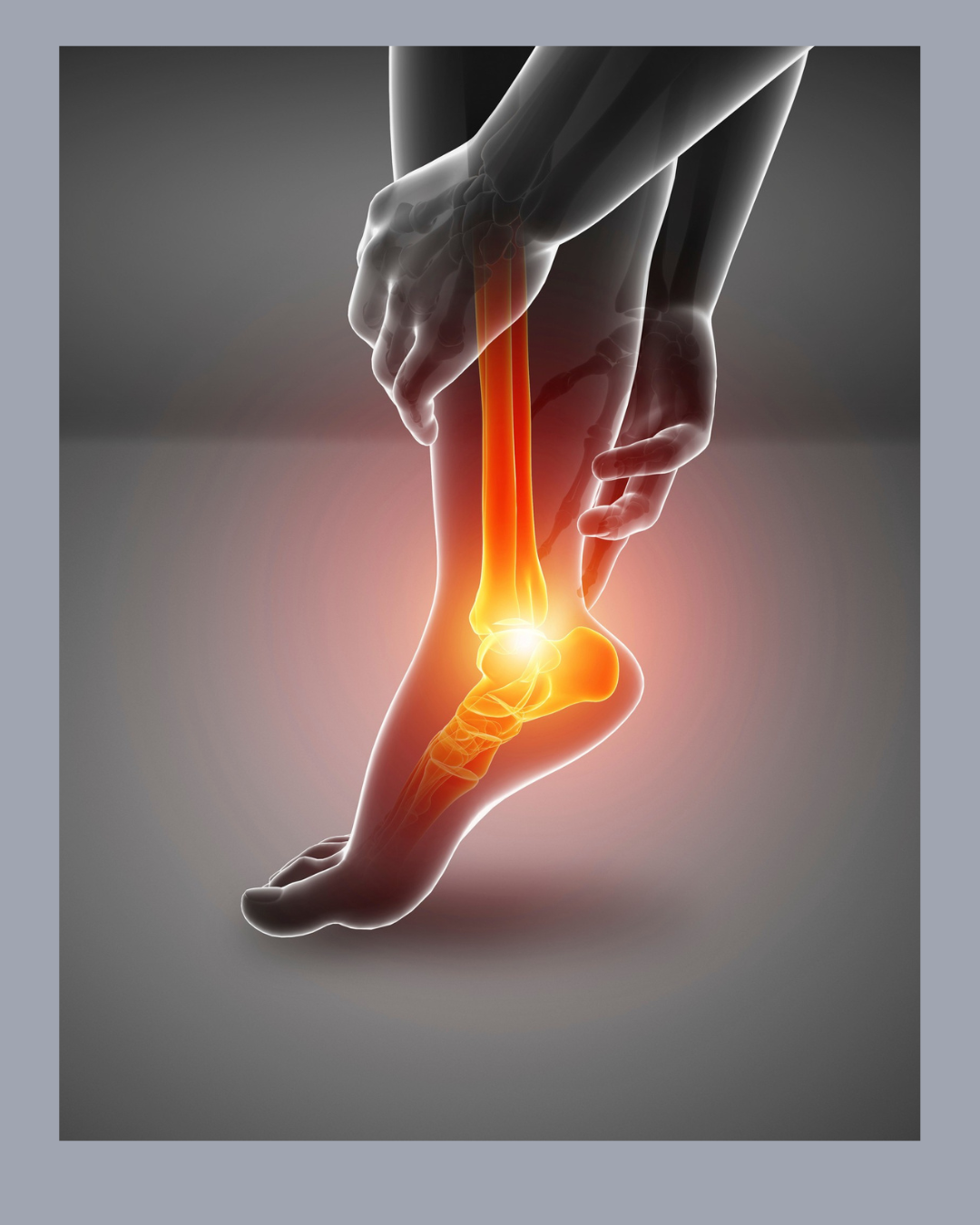
As a result, MRI is particularly beneficial in identifying conditions such as ligament tears, muscle strains, and tendon injuries that may not be detected by other imaging technologies.
These high-resolution images enable physiotherapists to accurately assess the extent of soft tissue damage, facilitating targeted treatment plans for patients seeking relief from pain and improved mobility.
Recommended for diagnosing issues with the brain, spinal cord, and joints
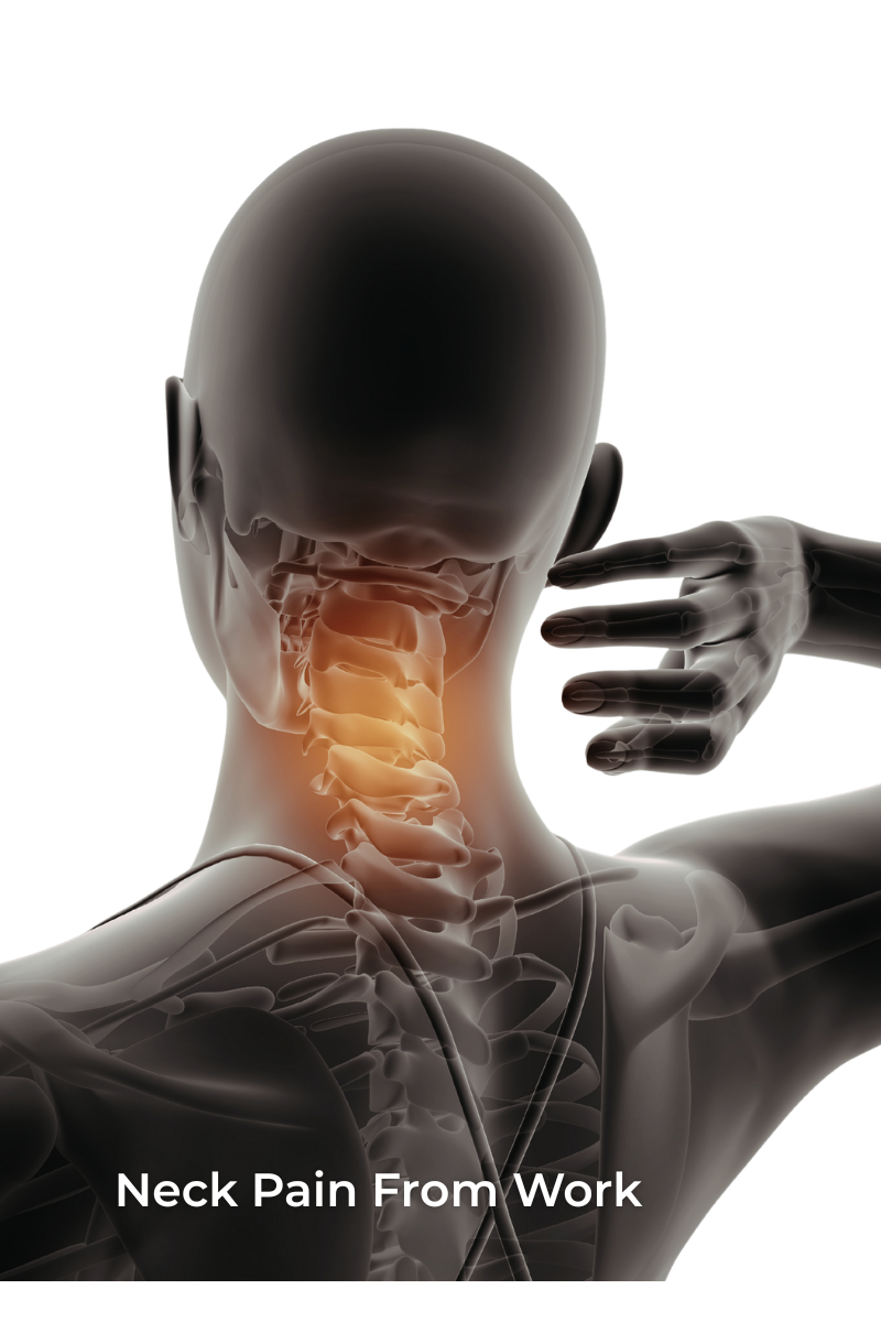
MRI is the preferred imaging option for detecting issues in the brain, spinal cord, and joints. This radiation-free technology provides detailed images of soft tissues, making it ideal for identifying injuries or diseases in these areas.
By utilising MRI scans, healthcare professionals can accurately diagnose conditions such as tumours, nerve damage, and joint disorders.
Physiotherapists may recommend an MRI to assess the extent of injury to the brain, spinal cord or joints so that appropriate treatment plans can be developed. This advanced imaging technique plays a crucial role in identifying underlying problems that may be causing pain or discomfort in these specific areas.
When to Choose an X-ray
X-rays are a quick and widely available imaging option used to detect fractures, dislocations, and other bone-related injuries. They are also recommended for diagnosing issues with the chest, abdomen, and skeletal system.
Quick and widely available imaging option
X-ray imaging is a widely available and rapid diagnostic tool. It can swiftly detect bone fractures, dislocations, and other skeletal injuries. X-rays are also useful for diagnosing issues related to the chest and abdomen.
This imaging technology is often readily accessible in medical facilities, making it an efficient option for promptly evaluating bone-related concerns.
Additionally, X-rays provide quick insights into skeletal conditions that may require immediate attention from a physiotherapist or other healthcare professionals focused on musculoskeletal health.
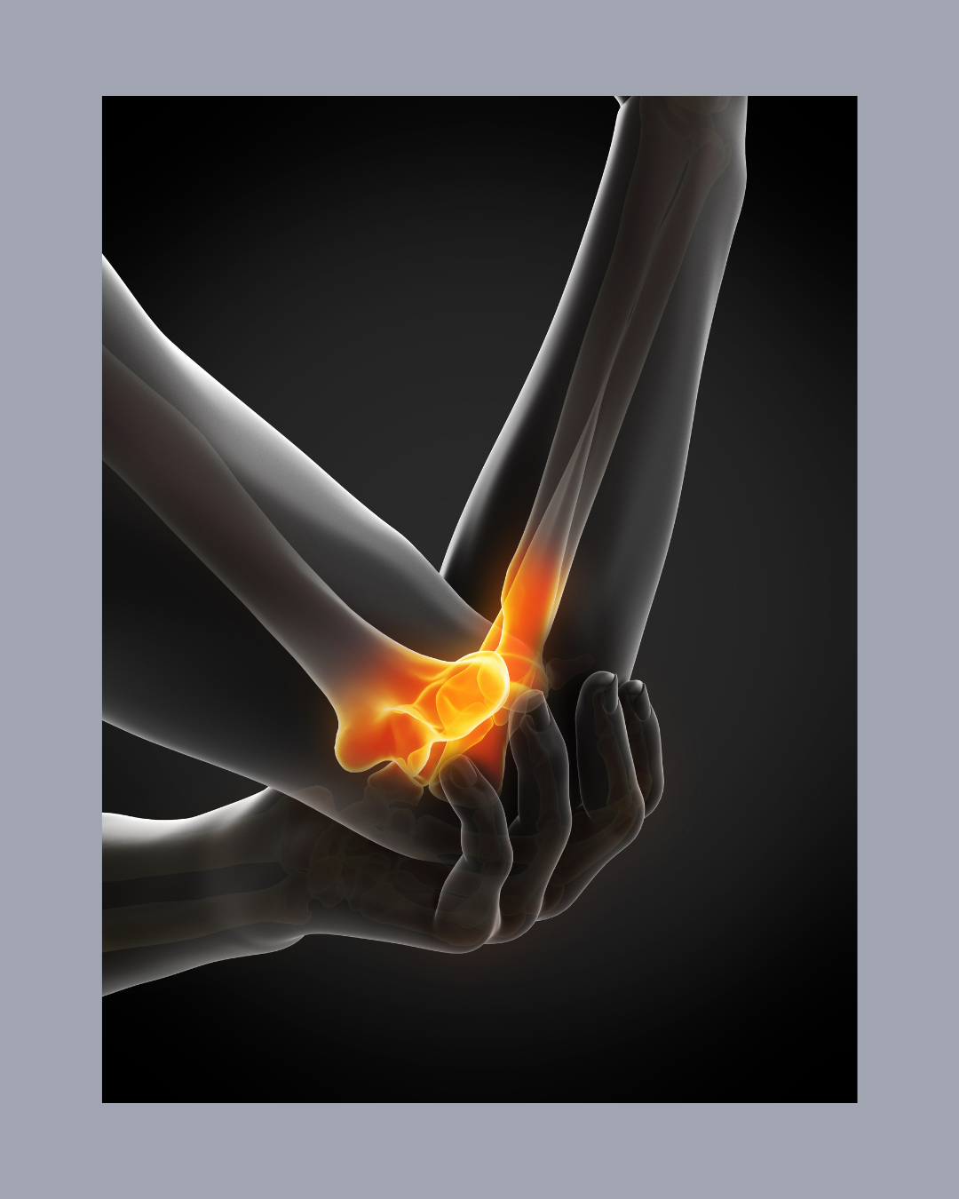
Used to detect fractures, dislocations, and other bone-related injuries
X-ray imaging is an essential tool for diagnosing fractures, dislocations, and other bone-related injuries. This quick and widely available imaging option allows healthcare professionals to obtain detailed images of the skeletal system, aiding in accurate diagnosis and treatment planning.
Physiotherapists often rely on X-rays to identify specific areas of concern within the bones, providing valuable insights for developing targeted rehabilitation plans.
Furthermore, X-rays play a crucial role in detecting abnormalities within the chest and abdomen. By capturing precise images of these areas, medical professionals can swiftly diagnose conditions that may impact an individual's overall health and well-being.
Recommended for diagnosing issues with the chest, abdomen, and skeletal system
X-ray imaging is recommended for diagnosing issues with the chest, abdomen, and skeletal system. It provides quick and widely available imaging for detecting fractures, dislocations, and other bone-related injuries.
This imaging technique is commonly used in medical diagnostics to assess conditions affecting the chest, such as pneumonia or lung cancer. In addition to bone injuries, X-rays are also employed to examine the abdominal area for potential issues in organs like the liver or intestines.
They play a crucial role in identifying skeletal abnormalities or structural irregularities that may require medical attention.
Conclusion
When determining the need for an MRI or X-ray, it's crucial to consider the specific diagnostic requirements. The MRI offers detailed soft tissue images and is a safe option without radiation.
On the other hand, X-rays are quick and widely available, ideal for detecting bone-related injuries. Each imaging technique serves a unique purpose in diagnosing different medical conditions based on their strengths and limitations.